What 'Best Ultrasound Scan Near Me' Actually Means — And How to Find It
10 Sep, 2026
21 Views 0 Like(s)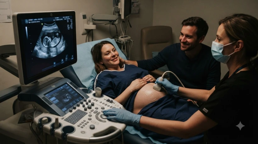
If you are looking in Bangalore, you can evaluate the best ultrasound scan near me at Ecotown Diagnostics, where modern equipment and experienced sonographers are part of the standard service.
Searching for the best ultrasound scan near me reflects something specific: you do not just want any facility that owns a machine. You want a scan that is conducted properly, interpreted accurately, and reported clearly. That is a higher and more meaningful standard than simply finding the closest available option.
This article explains what genuinely defines a high-quality ultrasound experience, what to check before booking, and what to expect during and after the scan. If you are looking in Bangalore, you can evaluate the best ultrasound scan near me at Ecotown Diagnostics, where modern equipment and experienced sonographers are part of the standard service.
Why Ultrasound Quality Varies So Much
Ultrasound is a skill-dependent imaging modality. Unlike a blood test where a machine processes the sample and produces a result, ultrasound requires a trained operator to use a probe, interpret real-time images, and recognise clinically relevant findings as they appear on screen.
This means two centres with similar machines can produce dramatically different diagnostic outcomes depending on the experience of the person performing the scan.
Factors that affect ultrasound quality:
Sonographer Experience
An experienced sonographer knows how to position the probe, adjust gain and depth settings, and identify structures that may be subtle, deep, or difficult to visualise. They know when a finding is incidental and when it warrants documentation and follow-up.
A less experienced operator may miss findings or misidentify normal anatomy as pathological — both carry clinical consequences.
Machine Specification
Higher-resolution ultrasound machines produce clearer images with greater tissue differentiation. Modern systems include features such as harmonic imaging, elastography, and Doppler modes that expand diagnostic capability beyond basic grey-scale imaging.
A low-specification machine limits what can be seen, regardless of operator skill.
Radiologist Involvement in Reporting
In some facilities, the sonographer performs the scan and produces a report. In others, a radiologist or specialist physician reviews the images and generates the official clinical report. The latter represents a higher standard of diagnostic quality.
Types of Ultrasound Scans and What Each Involves
Understanding which scan your doctor has prescribed helps you evaluate whether a particular facility is genuinely equipped for it.
Abdominal Ultrasound: Evaluates the liver, gallbladder, pancreas, spleen, and kidneys. One of the most common ultrasound investigations. Typically requires fasting for four to six hours beforehand to ensure the gallbladder is distended and bowel gas is minimised.
Pelvic Ultrasound (Transabdominal and/or Transvaginal): Evaluates the uterus, ovaries, and pelvic structures. A full bladder is required for transabdominal pelvic scans. Transvaginal ultrasound provides higher resolution pelvic detail and is performed with an empty bladder.
Obstetric Ultrasound: Pregnancy-related scanning at various gestational stages — first trimester dating and nuchal translucency, anomaly scans, growth scans, and Doppler studies. These require a sonographer with specific obstetric training.
Thyroid Ultrasound: Evaluates the thyroid gland and adjacent cervical lymph nodes. High-frequency probes are essential for adequate resolution of the small structures involved.
Scrotal Ultrasound: Assesses testicular structure, epididymis, and blood flow. Doppler capability is important for evaluating testicular torsion or varicocele.
Musculoskeletal Ultrasound: Evaluates tendons, ligaments, muscles, and joints. Requires a high-frequency probe and a sonographer with specific musculoskeletal training.
Doppler Ultrasound: Evaluates blood flow in arteries and veins — commonly used for deep vein thrombosis, carotid artery stenosis, and peripheral arterial disease.
When you book, confirm the facility regularly performs your specific scan type. Not all centres have equal experience across all ultrasound subspecialties.
Preparation: Getting It Right Before Your Appointment
Proper preparation is not just administrative box-ticking — it directly affects image quality.
|
Scan Type |
Preparation Required |
|
Abdominal |
4–6 hours fasting |
|
Pelvic (transabdominal) |
Full bladder (drink 1 litre of water, do not empty) |
|
Pelvic (transvaginal) |
Empty bladder |
|
Obstetric (first trimester) |
Full bladder |
|
Obstetric (second/third trimester) |
No special preparation usually |
|
Thyroid |
No preparation needed |
|
Renal / Urinary tract |
Adequate hydration |
|
Scrotal |
No preparation needed |
Confirm preparation requirements with the centre when booking, as protocols can vary slightly. Arriving without the correct preparation may mean a rescheduled appointment.
What to Expect During the Scan
Most ultrasound appointments follow a predictable sequence.
-
You will be positioned on a couch — lying flat or slightly elevated depending on the scan area.
-
A water-based gel is applied to the skin surface. This gel conducts sound waves and eliminates air between the probe and the skin.
-
The sonographer moves the probe across the relevant area, capturing images in real time.
-
You may be asked to breathe in, breathe out, or hold your breath briefly to optimise visualisation of specific structures.
-
The entire scan typically takes fifteen to forty-five minutes, depending on the study complexity.
Ultrasound is completely painless and uses no radiation. It is safe for all patient groups, including pregnant women and children.
Getting the Most from Your Report
A well-written ultrasound report describes the structures examined, their size, echogenicity, and any abnormalities detected. It should answer the specific clinical question raised in your referral.
When you collect your report:
-
Read through the findings section, even if the terminology is unfamiliar
-
Note any terms like "further evaluation recommended" — this needs prompt follow-up with your doctor
-
Bring both the written report and any saved images (if provided on CD or digitally) to your follow-up appointment
If you have follow-up questions about what the report means clinically, your doctor is the right person to answer them in the context of your overall health picture.
Frequently Asked Questions
What makes one ultrasound centre better than another?
The key differentiators are sonographer experience, machine quality, and whether a qualified radiologist or specialist physician reviews and reports the findings. A centre where all three are strong produces consistently reliable diagnostic output.
Is it always necessary to fast before an ultrasound?
Fasting is specifically required for abdominal ultrasound involving the gallbladder and upper abdominal organs. It is not required for thyroid, scrotal, obstetric, or musculoskeletal ultrasounds. Your centre will advise what applies to your specific scan.
Can ultrasound detect cancer?
Ultrasound can identify suspicious masses, unusual tissue characteristics, and abnormal lymph nodes that may prompt further investigation. However, it cannot confirm or exclude cancer on its own — further tests such as biopsy or MRI are typically required for a definitive diagnosis.
How soon will my ultrasound report be ready?
At most well-run facilities, reports are available the same day or within a few hours of the scan. Some complex or specialist studies may take one business day. Confirm the expected timeline when you book.
Is ultrasound safe during pregnancy?
Yes. Ultrasound uses sound waves, not ionising radiation, and has an established safety record throughout all stages of pregnancy. It is the primary imaging modality used in obstetric care precisely because of this safety profile.
What if the sonographer finds something unexpected during my scan?
If an unexpected finding is identified during the scan, the sonographer or radiologist will document it in the report. Your doctor will then advise on appropriate next steps — whether that is further imaging, a specialist referral, or watchful monitoring. It does not mean the worst-case scenario; many incidental findings are benign.
The best ultrasound scan near me is not simply the nearest option — it is the one where trained hands operate quality equipment and produce a report your doctor can act on with confidence. These are achievable standards at well-run diagnostic centres in Bangalore. Spending a few minutes confirming them before booking is the simplest way to ensure your scan genuinely serves its diagnostic purpose.
Comments
Login to Comment