Can an Echo Test in Bangalore Detect Blockages in Your Heart? What Most Patients Get Wrong
10 Sep, 2026
3 Views 0 Like(s)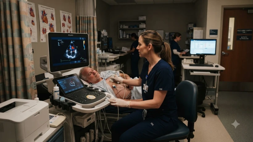
The reality is more nuanced. An echo test in Bangalore can tell your doctor a great deal about the heart’s structure and function, but it cannot always tell you whether the coronary arteries are narrowed or blocked.
It is one of the most common assumptions made in cardiac diagnostics: “I had an echo test, so my heart is clear.” Patients come out of an echocardiogram scanning centre believing that they have been checked for a heart attack risk, blocked arteries, and the need for angioplasty, all in one 30-minute ultrasound.
The reality is more nuanced. An echo test in Bangalore can tell your doctor a great deal about the heart’s structure and function, but it cannot always tell you whether the coronary arteries are narrowed or blocked. Understanding this distinction can prevent false reassurance on the one hand and unnecessary panic on the other.
Why Patients Assume an Echo Can See Blocked Arteries
The word “blockage” causes most people to think of circulatory plumbing. Since the heart is a muscle that needs blood supply, it seems logical that an imaging test would be able to see pipes and narrowing.
But echocardiography images the heart walls, chambers, and valves. The coronary arteries, which deliver oxygen-rich blood to the heart muscle itself, are small, curved vessels that move with every heartbeat. Routine transthoracic echo usually does not visualise them clearly enough to measure stenosis.
That is why an entirely normal echo report does not mean your arteries are free of disease. It only means that the heart muscle, valves, and chambers look structurally and functionally normal at the time of the test.
What an Echo Test Can Actually Detect
Even though it cannot show coronary blockages directly, echocardiography is a powerful diagnostic tool. It can identify:
-
Weakened heart muscle function. The report usually gives an estimation of ejection fraction, which tells your doctor how effectively your heart is pumping blood out to the body.
-
Chamber enlargement. Chronic high blood pressure or valve disease can cause the heart chambers to stretch or thicken.
-
Valve abnormalities. The echo can show whether valves are narrowed, leaking, or failing to close properly.
-
Heart wall motion abnormalities. If a part of the heart muscle was previously damaged by a heart attack, it may not move as vigorously as the rest of the heart.
-
Fluid around the heart. Excess fluid in the pericardial sac can compress the heart and impair its function.
-
Congenital structural defects. Abnormal connections between chambers or vessels can be seen in many cases.
This is why an echo is so useful. It gives the doctor a real-time view of the heart in motion, rather than just an electrical tracing on an ECG.
What It Cannot Detect
Echocardiography has limitations that every patient should understand:
-
Early coronary artery blockages. Small plaques that narrow an artery but have not yet damaged the heart muscle are usually invisible on a resting echo.
-
Electrical rhythm problems. An echo is not the right test to diagnose arrhythmias. That requires an ECG or Holter monitor.
-
The exact cause of chest pain. Multiple conditions can cause chest discomfort. An echo helps with some of them, but not all.
If a blockage is severe enough to cause a recent heart attack, the affected heart wall may move abnormally. That movement abnormality can sometimes be detected on echo. But a mild or even moderate narrowing in a coronary artery may not affect the resting heart wall motion at all.
Which Tests Are Used Alongside an Echo?
When your doctor suspects coronary artery disease, the echo is rarely the final test. It is often combined with:
-
An ECG to look for electrical signs of previous or ongoing heart muscle damage
-
A treadmill stress test (TMT) to see whether the heart develops breathing problems during exercise
-
A stress echo to compare heart wall motion at rest and immediately after stress
-
A CT coronary calcium score to estimate plaque burden in the arteries
-
A CT coronary angiogram to directly visualise coronary blockages
-
Cardiac catheterisation if a significant blockage is strongly suspected
Each test answers a different question. The echo asks: “What does the heart look like and how well is it pumping?” The TMT asks: “Does the heart receive enough blood supply when it works harder?”
What to Do If You Still Have Symptoms With a Normal Echo
You can experience chest pain, discomfort, or palpitations and still have a normal resting echo. In fact, this is common with exertional angina.
If your doctor referred you for an echo test in bangalore as part of a cardiac workup, but your symptoms continue, you must not interpret a normal report as proof that nothing is wrong. Go back to your doctor and explain that your symptoms persist.
Doctors may recommend an exercise stress test, ambulatory ECG monitoring, or further imaging depending on the pattern of your symptoms. Risk factors such as family history, diabetes, smoking, high cholesterol, and high blood pressure will also guide the next step.
What Do the Terms in Your Echo Report Mean?
Most echo reports contain terms that patients try to interpret at home. A few common examples:
-
Ejection fraction (EF). This is a percentage representing how much blood the left ventricle pumps out with each heartbeat. It is one of the most widely used measures of heart failure.
-
Wall motion abnormalities. Segments of the heart muscle that do not contract normally.
-
Regurgitation. Backward leakage of blood through a heart valve. Trace or mild regurgitation is common and often benign.
-
Stenosis. A valve that does not open fully, restricting forward blood flow.
Only a doctor who knows your clinical history can translate these terms into a clear treatment plan.
When an Echo Is the Right Choice
An echo is an excellent test in many situations:
-
To evaluate a heart murmur
-
To check heart function in a person with breathlessness or leg swelling
-
To monitor heart function after a heart attack
-
To follow patients who are receiving certain chemotherapy medicines
-
To assess structural heart disease in those with high blood pressure
In all of these situations, the question is about the heart muscle, chambers, or valves, not primarily about the coronary arteries.
Frequently Asked Questions
Is an echo test in bangalore enough to rule out a heart attack risk?
No. It evaluates structure and pumping function, but it does not directly measure plaque in the coronary arteries. Your overall heart attack risk depends on many factors, including age, blood pressure, cholesterol, diabetes, and smoking history.
Can an echo be done during a heart attack?
Yes. A bedside echo is sometimes used during an emergency heart attack to assess how much heart muscle is damaged and to detect complications. It does not replace the ECG or blood tests used to diagnose a heart attack.
Will I feel anything during an echo?
Only pressure from the ultrasound probe on your chest. There are no needles, no dyes, and no radiation. The main discomfort comes from lying on your side and holding your breath at various points during the scan.
If your doctor sends you for an echo test in Bangalore to check your symptoms, ask what specific question the test is meant to answer. That one question will help you understand whether your results, normal or abnormal, are truly the final word or just one piece of a larger cardiac picture.
Comments
Login to Comment