Can Artificial Intelligence Detect Retinal Damage Before Vision Changes
15 Oct, 2026
8 Views 0 Like(s)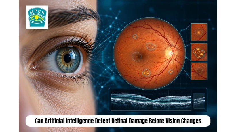
Can AI spot retinal damage before vision changes? Learn when to see a retina specialist in Ranchi.
You squint at a road sign, and it looks fine. You read your phone, and that looks fine too. Meanwhile, tiny vessels inside your eye may already be leaking, and your brain is too polite to mention it. Retinal damage rarely announces itself. By the time blurry patches, dark spots or floaters appear, the injury has often run for months or years.
That delay is frightening because lost retinal tissue rarely grows back. India feels this pressure sharply. A global diabetes atlas for 2025 reports that 89.8 million Indian adults lived with diabetes in 2024, and many of them do not know it. So can artificial intelligence spot trouble before your vision changes? For diabetic eye screening, it increasingly can.
This guide explains how algorithms read eye images, what a 2026 systematic review found, where research ends and daily practice begins, and why a trusted retina specialist in Ranchi still holds the final word. Keep reading to learn what to expect and when to act.
Why Retinal Damage Stays Silent Until It Is Late
The retina rarely complains while it is being hurt. This section explains how the damage builds, why you cannot feel it and why India's numbers make early checks so urgent.
How Diabetes Quietly Harms the Retina
The retina works like the film in an old camera. It captures light and sends a picture to the brain. Damage here behaves like a quiet roommate who never complains. Early diabetic retinopathy causes no pain, no redness and often no blurring. High blood sugar weakens tiny vessels. They bulge, leak fluid and sometimes close off. Fluid collecting at the centre produces macular oedema, and that is when reading suddenly becomes a struggle.
Many readers ask, can retinal damage happen without any symptoms? Yes, and that is exactly where the problem lies. Central vision may stay sharp while damage creeps through the surrounding tissue. Your brain also blends the view from both eyes, which hides early vision loss.
Why Millions Miss the Warning Window
India's 2025 national guidelines estimate that around 101 million adults live with diabetes. Roughly 5 to 7%, about 4 to 6 million people, already face sight-threatening diabetic retinopathy. The atlas also notes that 43% of Indian adults with diabetes do not know they have it. Nobody books a dilated eye exam for a disease they have never heard they have.
People often wonder, How frequently should people with diabetes undergo retinal examinations? Most guidelines advise an eye examination at diagnosis for type 2 diabetes, then at least once every year. Your doctor may shorten that gap if retinopathy already exists.
How AI Reads Fundus Photographs, OCT Scans and OCT Angiography
An algorithm cannot examine a patient. It examines images, and it does so very fast. Three image types dominate current research, and each one shows a different layer of the story.
Fundus Photographs
A fundus photograph is a colour picture of the back of the eye. A camera captures it in seconds. Deep learning models, especially convolutional neural networks, scan every pixel for microaneurysms, haemorrhages and hard exudates. A comprehensive review of 91 studies found that 81% used fundus images, which makes this the most mature approach.
OCT Scans
Optical coherence tomography works like an ultrasound that uses light. It builds cross sections of the retina and measures retinal thickness with remarkable precision. AI studies this layered view to spot swelling, fluid pockets and thinning that a flat photograph can miss. Only 9% of the reviewed studies used OCT, so this area still has room to grow.
OCT Angiography
OCT angiography, often called OCTA, maps blood flow without dye injections. Curious patients ask, what can OCT angiography see that a normal eye photo cannot? It reveals capillary dropout, an early microvascular change that ordinary photographs may not show. About 6% of reviewed studies used it. Models learn to grade these flow maps, which makes OCTA a promising route toward early detection.
What the 2026 Evidence Actually Says
Research on this topic has exploded. This section sums up the biggest reviews and explains how far their findings can travel into real clinics.
Scale of the Research
A 2026 systematic review searched five major databases up to March 2026. It identified 1,007 articles and kept 175 studies that applied AI to OCT and OCTA images for diabetic retinopathy. That volume shows how fast the field moves. Convolutional neural networks and newer attention-based models dominate the work.
|
Evidence |
Scale |
Main message |
|
2026 review of OCT and OCTA |
175 studies included |
Deep learning dominates analysis of structural and vascular retinal images |
|
2025 meta-analysis |
47 studies, 188,268 images and scans |
Deep learning shows high accuracy for detecting diabetic retinopathy |
|
Broad review of AI grading |
91 studies, 23 public datasets |
Deep learning generally beats older methods across image types |
Why Big Numbers Do Not Guarantee Clinic Success
Big numbers tempt people to cheer early. Readers rightly ask, does a large number of studies prove AI works in every clinic? No. Most studies train and test on curated image collections. Real clinics bring blurry images, small pupils and tired patients. A model that shines on a tidy dataset may stumble in a busy hospital room, so local validation matters as much as published accuracy.
Which Retinal Problems AI Can Flag Early
AI does not treat every eye disease equally well. This section separates the conditions with strong evidence from those where the science is still young.
Diabetic Retinopathy
Diabetic retinopathy leads the list because the evidence is deepest. A pivotal trial of the first autonomous system cleared by a United States regulator in 2018 reported 87.2% sensitivity and 90.7% specificity for more than mild disease. Sensitivity tells you how many sick eyes the system catches. Specificity tells you how many healthy eyes it leaves alone. A good screening tool needs both; otherwise it either misses patients or floods clinics with false alarms. Patients frequently search for: How accurate is AI in detecting diabetic retinopathy? Published results are strong, yet accuracy depends on image quality, patient mix and the device used.
Macular Oedema and Retinal Vascular Abnormalities
OCT-based models measure fluid and thickness at the macula, which helps flag macular oedema before central vision drops. Other models study vessel patterns for retinal vascular abnormalities, including blocked branches and changes linked to high blood pressure. Researchers also test AI for age-related macular degeneration and retinal detachment risk. The evidence here is thinner than for diabetic retinopathy, so doctors treat these outputs as helpful hints, not final answers. A flagged scan simply earns a faster look from an ophthalmologist and a quicker referable retinopathy decision.
Research Promise Versus Everyday Clinical Practice
Headlines often blur the line between a lab result and a bedside tool. This section shows which applications already help patients and which still wait for proof.
Where AI Stands Today
A simple comparison keeps expectations honest.
|
Application |
Current status |
Realistic role |
|
Fundus photo screening for diabetic retinopathy |
Established in some settings, with cleared autonomous systems |
Screening aid that triggers referral |
|
OCT fluid and thickness analysis |
Growing, often built into scanners |
Decision support for the doctor |
|
OCTA-based staging |
Mostly research |
Promising, needs larger validation |
|
Predicting future progression |
Early research |
Not ready for routine decisions |
How AI and Doctors Work Together
Many people ask, is AI already used in hospitals to detect retinal disease? Yes, mainly for diabetic retinopathy screening and as software inside retinal imaging devices. Many other uses remain experimental. The honest summary says AI works best as a fast, tireless first reader. It never examines your whole eye, asks about your symptoms or weighs your blood sugar history. Your doctor does all of that, and the best results come when both work together.
Limits and Risks Every Patient Should Know
AI carries real weaknesses, and good clinics talk about them openly. This section lists the main risks and explains why human oversight stays essential.
Key Weaknesses of AI Screening
Consider these points before trusting any automated result.
- Biased data. Models trained on one population may stumble on another with different retinal pigmentation or disease patterns.
- Poor image quality. Cataracts, small pupils and motion blur can fool even strong algorithms.
- False negatives. A missed case can create false comfort, which is the exact danger screening aims to avoid.
- False positives. Extra referrals cost time and worry.
- Black box reasoning. Many models cannot explain why they flagged an eye.
- Missing care pathways. A flagged image helps nobody unless treatment follows quickly.
Why Human Specialists Stay Essential
A common worry is, will AI replace my retina specialist? No. AI reads images, but a specialist reads the patient. Treatment decisions, including laser therapy, intravitreal injections and vitrectomy, need human judgment, consent and surgical skill. Think of AI as a smoke detector. It warns you loudly, but it never puts out the fire.
Where Retinal Imaging Meets Expert Judgment in Ranchi
Good technology still needs a good team behind it. This section shows what to look for in local retinal care and how to reach it.
Retinal Care Available at MPEH
Families searching for the best eye hospital in Ranchi should look past buzzwords and ask what imaging the team actually uses. Matri Prerna Multispeciality Eye Hospital (MPEH) in Kokar, Ranchi, presents itself as a leading, nationally accredited eye hospital. Its retinal care covers diabetic retinopathy, age-related macular degeneration, retinal detachment, macular holes, vascular occlusions and hypertensive retinopathy. The hospital states that it uses OCT scans and angiography for diagnosis, along with laser therapy, vitrectomy and injections for treatment.
Many patients ask, what should I look for in a retina specialist in Ranchi? Look for modern imaging, clear explanations and an ophthalmologist who interprets every scan personally. Imaging supplies the same kind of data that AI research studies, but only an experienced doctor turns it into a plan.
Symptoms That Need a Quick Visit and Booking Details
See a doctor quickly if you notice blurred or distorted vision, sudden flashes, new floaters, dark spots or lost side vision. Individuals with diabetes or high blood pressure should also schedule regular eye examinations without waiting for symptoms to appear.
The hospital runs Monday to Saturday, 9 AM to 6 PM. Call +91 7541825700 or 0651 2544676. MPEH accepts in-person appointments only, so avoid anyone who charges you for online booking.
Conclusion
Artificial intelligence can read fundus photographs, OCT scans and OCT angiography images faster than any human team, and it can flag signs of diabetic retinopathy and macular oedema before you notice a single symptom. The 2026 systematic review of 175 studies proves the field is booming, and earlier meta-analyses confirm high accuracy under study conditions.
Even so, strong research is different from routine practice. Fundus-based diabetic retinopathy screening has the firmest footing. OCT analysis is growing steadily, while OCTA staging and progression prediction still need larger validation.
AI also has limits. Biased data, poor image quality and missed cases keep human oversight essential. India's diabetes numbers make early eye checks urgent, because millions live with risk they cannot feel. Do not wait for blurry vision to start the conversation. Book a retinal examination, ask which imaging tools the clinic uses and make sure an experienced ophthalmologist reviews every result. Your eyes will not send a warning letter, so schedule the visit yourself.
Frequently Asked Questions (FAQs)
1. Can AI detect diabetic retinopathy before I notice vision problems?
Yes. AI analyses retinal images for microaneurysms, leakage and swelling that cause no symptoms early on. It works as a screening aid, and a doctor must confirm any flagged finding.
2. How frequently should someone with diabetes have their retina examined?
Most guidelines advise an eye examination at diagnosis for type 2 diabetes, then at least once every year. Your doctor may recommend shorter gaps if retinopathy already exists.
3. Is an AI eye screening as reliable as an ophthalmologist's examination?
AI screening performs well for referable diabetic retinopathy, yet it checks only images. An ophthalmologist examines the whole eye, reviews your history and decides treatment, so AI supports rather than replaces that visit.
4. How does OCT differ from OCT angiography?
OCT shows retinal layers and thickness, which helps detect swelling and fluid. OCT angiography adds a map of blood flow in tiny vessels without dye, revealing circulation problems earlier.
5. When should I visit a retina specialist in Ranchi?
Visit promptly if you see flashes, new floaters, dark patches, distorted lines or lost side vision. People with diabetes or high blood pressure should also schedule regular eye examinations rather than wait for symptoms to appear.



Comments
Login to Comment